Bladder scanner
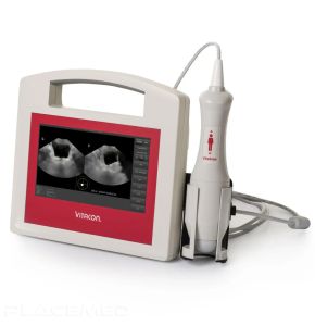
Portable Ultrasound Bladder Scanner - VitaScan PD 100570CP
Price on requestBladder scanner Certified professional
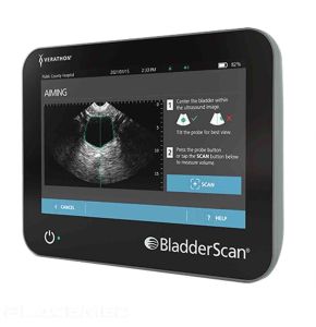
The BladderScan i10 bladder scanner by VERATHON
Price on requestBladder scanner Certified professional
Discover the ultimate precision in bladder imaging with our dedicated "Bladder Scanner" booth: a wide range of bladder scanners are exhibited by our professional partners. Our rigorous selection offers the promise of cutting-edge technology for precise and fast diagnosis.
From detecting bladder abnormalities to monitoring treatments, these medical tools are designed to optimize patient care while ensuring easy use and long-lasting performance. Take a step towards medical excellence - explore our booth today and choose from the best equipment in the medical device market.
What is a BLADDER SCANNER?
A bladder scanner, also known as a bladder ultrasound, is a high-tech medical tool used to create detailed images of the bladder and surrounding structures. It uses ultrasound technology or sound waves to produce these images, providing doctors with a clear and precise view of the area, thus aiding in the diagnosis, monitoring, and treatment of various medical conditions.
This medical device plays a crucial role in medical diagnosis, particularly in detecting bladder abnormalities such as tumors, bladder stones, infections, or inflammatory conditions. It also helps assess the bladder's capacity to store and release urine, which can aid in identifying the causes of urinary incontinence or urinary retention.
By offering real-time and non-invasive visualization of the bladder, the bladder scanner greatly contributes to improving diagnostic accuracy and selecting the most appropriate treatment for the patient. Moreover, it can also be used to monitor the effectiveness of ongoing treatments, making it an indispensable tool in modern medical practice.
What are the different types of BLADDER SCANNERS?
The field of bladder medical imaging mainly comprises two types of bladder scanners:
Ultrasound Bladder Scanner
This is the most commonly used type for bladder imaging. It uses high-frequency sound waves that bounce off tissues and organs to produce an image. It is non-invasive, radiation-free, and provides a good representation of the bladder's structure and surrounding organs.
Bladder Scanner by Computerized Tomography (CT)
This type of scanner uses X-rays to create a detailed image of the bladder and surrounding organs. It can provide more detailed images than ultrasound, especially for visualizing small tumors or bladder stones. However, due to the use of X-rays, it needs to be used with caution to minimize radiation exposure.
Principle of Operation of a BLADDER SCANNER
This type of equipment allows healthcare professionals to obtain detailed images of the bladder and surrounding structures, thus diagnosing, monitoring, and treating a multitude of medical conditions. But how does it exactly work, and what are the different technologies involved?
Indeed, its operation depends on the technology used. Generally, the patient is positioned in a way that allows the device to be directed towards the bladder. For ultrasound scanners, a gel is applied to the skin to aid in the transmission of sound waves. A probe then emits sound waves that bounce off the tissues and are captured by the same probe. The device's software then interprets these echoes to produce an image of the bladder. For a CT-type scanner, the patient lies on a table that slowly passes through a ring of X-rays. Detectors record the amount of X-rays (or photons) that pass through the body, and this information is used to generate an image.
As for the technologies used in these types of devices, there are primarily two types: ultrasound and computerized tomography (CT). Ultrasound is commonly used due to its non-invasive nature and lack of radiation. It is particularly effective in visualizing the structure of the bladder and surrounding soft tissues. CT scanners, on the other hand, offer higher resolution and are particularly useful for visualizing small anomalies such as tumors or bladder stones. However, they involve some radiation exposure, so they need to be used with caution.
Use of a BLADDER SCANNER
The use of this type of imaging device in medicine plays a crucial role in the diagnosis and monitoring of various medical conditions affecting the bladder. But when is it used, how should one prepare, and what happens during a session?
Indications for Use
This type of device is generally used when symptoms such as pelvic pain, frequent urination, or blood in the urine suggest a bladder abnormality. It is also used to monitor the treatment of bladder diseases, such as bladder cancer. Additionally, it can be used to evaluate the condition of the bladder in conditions such as urinary incontinence or urinary retention.
Preparation
The preparation may vary depending on the type of device. For an ultrasound scanner, the patient is usually instructed to drink plenty of water before the examination to fill the bladder, which improves the image quality. For a CT scanner, a contrast solution may be administered orally or intravenously to help distinguish different structures. It is always advisable to follow the specific instructions provided by your doctor or imaging technician.
Procedure
During a bladder scanner session, the patient is typically positioned lying on an examination table. For an ultrasound scanner, a gel-coated probe is moved over the pelvic area to obtain the images. For a CT scanner, the examination table moves slowly through the machine's ring while images are taken. A bladder scanner is generally quick and painless, although the patient may experience some discomfort due to the pressure from the probe or examination table.
Advantages and Disadvantages of Bladder Scanning
Bladder scanning, although widely used in medical practice, has both advantages and disadvantages, and its performance can be compared to other medical imaging techniques.
Advantages
One of its main advantages is its ability to provide detailed images of the bladder and surrounding organs, aiding in the accurate diagnosis of various medical conditions. Additionally, it is non-invasive and generally painless. Compared to other imaging techniques, it can offer better visualization of certain abnormalities, such as small tumors or bladder stones. Furthermore, monitoring changes over time is facilitated by the ability to compare current images with those from previous examinations.
Limitations
However, bladder scanning also has its limitations. For example, radiation exposure with CT scanners may be a concern, although the dose used is typically low. Additionally, some patients may have an allergic reaction to the contrast solution used in a CT scan. Furthermore, image quality may be affected in obese patients or those who cannot remain still during the examination.
Comparison with Other Medical Imaging Techniques
Compared to MRI, which can also provide detailed images of the bladder, bladder scanning is generally faster and less expensive. However, MRI may be preferred for certain conditions due to its superior resolution of soft tissues. Compared to cystoscopy, an invasive procedure involving the insertion of a thin tube into the urethra to visualize the bladder, bladder scanning is significantly more comfortable for the patient and carries fewer risks of complications.
How to Choose a Bladder Scanner?
Choosing an appropriate bladder scanner is a key decision for any clinic, hospital, or medical practice. Several factors need to be considered to ensure that the equipment meets the specific needs of the medical setting.
- Technology: Bladder scanners primarily use two types of technologies: ultrasound and computerized tomography (CT). Ultrasound scanners are non-invasive and radiation-free, while CT scanners offer more detailed images. The choice depends on the type of patients and the conditions commonly treated.
- Image quality: Clear and detailed imaging is essential for accurate diagnosis. Make sure to choose a scanner that provides high-quality image resolution.
- User-friendliness: The device should be user-friendly for the medical staff. User-friendly software, intuitive interface, and effective training capabilities are crucial.
- After-sales service and maintenance: Ensure that the provider offers good after-sales service, including maintenance and repairs when needed.
- Cost: Cost is also an important factor. This includes not only the initial cost of the device but also maintenance costs, necessary supplies costs, and staff training costs.
- Reviews and recommendations: Read reviews from other users and seek recommendations from colleagues or professional associations.
 Francais
 Quote
Quote  Cart
Cart 
