Microscope
18/11/2024 245
18/11/2024 248
18/11/2024 242
18/11/2024 248
18/11/2024 238
18/11/2024 314
18/11/2024 265
18/11/2024 245
18/11/2024 214
18/11/2024 243
18/11/2024 246
18/11/2024 271
Microscope: Serving Health by Exploring the Invisible
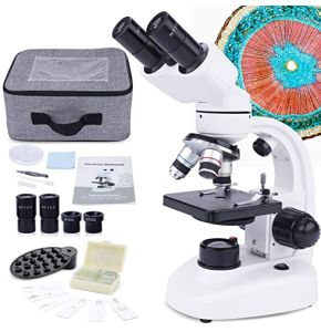
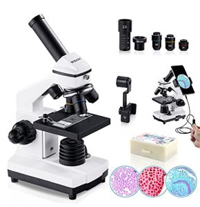
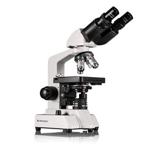
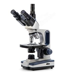
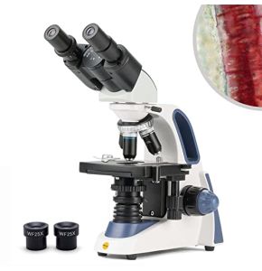
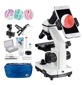
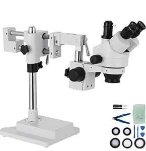
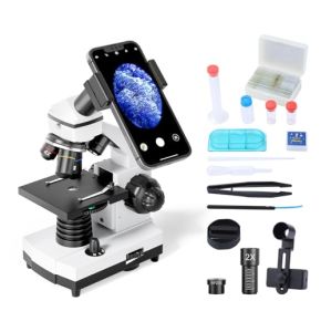
In this microscope category, Placemed offers you a complete range of high-quality microscopes to meet all your needs in the medical laboratory. Microscopes are essential instruments that allow scientists and doctors to explore the microscopic world. They play an important role in diagnosis, research, and education by revealing details invisible to the naked eye.
What is the Role of the Microscope in the Medical Laboratory?
It is a fundamental tool in medical laboratories. It allows the observation of cells, bacteria, viruses, and other microscopic structures. Thanks to it, healthcare professionals can:
- Diagnose Diseases: By examining tissue samples or bodily fluids.
- Identify Pathogens: Such as bacteria or viruses responsible for infections.
- Study Cell Structure: To understand their function and detect abnormalities.
Without the microscope, many advances in medicine would not be possible. It is indispensable for ensuring accurate diagnoses and appropriate treatments.
What is Its History?
Its history begins in the 16th century with the first magnifying glasses. But it was in the 17th century that the word microscope truly appeared. The Janssen brothers invented the compound model, which was a significant advancement. Subsequently, Antonie van Leeuwenhoek improved the lenses and created the simple model. These microscopes allowed the first-time viewing of small entities like cells and microorganisms. This opened new possibilities for scientific research. In the 20th century, the electron microscope was invented. It can magnify objects thousands of times more than optical microscopes. This revolutionized fields like biology and medicine. Today, modern techniques like fluorescence microscopy or atomic force microscopy allow the creation of three-dimensional images and the study of phenomena at the atomic level.
How Have Microscopic Technologies Evolved?
Since its invention, this medical device has undergone numerous improvements. The first microscopes used simple lenses to enlarge images. Today, advanced technologies offer better resolution and new observation possibilities.
Innovations include:
- Electron Microscopes: Which use electron beams to see details at the nanometer scale.
- Fluorescence Microscopes: Which allow the visualization of specific structures using fluorescent markers.
- Confocal Microscopes: Which provide three-dimensional images by scanning the sample at different depths.
These advancements have expanded the applications of the microscope and improved the quality of observations.
What Are the Different Types of Microscopy?
Optical Microscopy
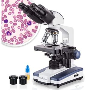
Optical microscopy is the most common form. It uses visible light and lenses to enlarge images. It is ideal for observing cells, tissues, and living organisms.
Electron Microscopy
Electron microscopy uses electrons instead of light. This allows for images with very high resolution. There are two main types:
- Transmission (TEM): For viewing the inside of cells.
- Scanning (SEM): For viewing the surface of samples in three dimensions.
Fluorescence Microscopy
This technique uses fluorescent dyes that emit light when illuminated. It allows the labeling and visualization of specific structures within cells.
Confocal Microscopy
Confocal microscopy uses lasers to obtain clear images at different depths. It allows the creation of three-dimensional images of the sample.
What Are the Specific Clinical Applications of Each Type?
Each type has its own uses:
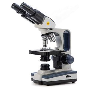
- Optical: General observation of cells and tissues, ideal for school and medical laboratories.
- Electron: Detailed study of structures at the atomic level, used in advanced research.
- Fluorescence: Visualization of specific molecules, useful in molecular biology and genetics.
- Confocal: Deep tissue analysis, employed in neuroscience and histology.
The choice of microscope depends on the type of sample and the information sought.
What Are the Technological Innovations in Microscopy?
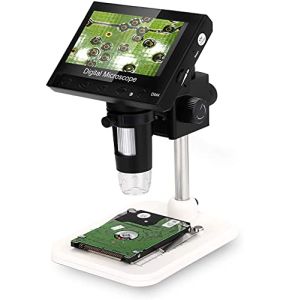
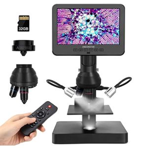
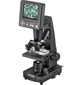
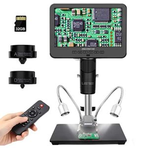
Digital Microscopes with Image Capture and Analysis
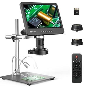
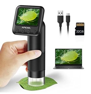
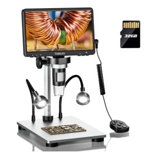
Digital microscopes integrate cameras to capture digital images. This allows:
- Storage: Record and archive images for later examination.
- Analysis: Use software to measure and interpret data.
- Sharing: Send images to other professionals for consultation.
Automation and Robotization of Analyses
Automation allows the processing of numerous samples quickly. Robotic systems can:
- Automatically change samples.
- Perform analyses without human intervention.
- Standardize procedures for greater reliability.
Image Sharing for Telepathology
Telepathology allows doctors to examine images remotely. This offers:
- Collaboration: Consult experts anywhere in the world.
- Speed: Obtain diagnoses more quickly.
- Teaching: Share cases for training purposes.
What Are Its Clinical Applications?
Histology Diagnosis
In histology, this device allows the examination of tissues to detect abnormalities such as cancers or inflammations.
Hematology Analysis
It is used to study blood cells, helping to diagnose diseases like anemia or leukemia.
Rapid Identification of Pathogens in Microbiology
In microbiology, the microscope helps identify bacteria and viruses responsible for infections, which is essential for choosing the right treatment.
How Do Training and Skills Evolve with New Technologies?
With the advent of new technologies, professionals must update their skills. This includes:
- Learning New Tools: Knowing how to use digital microscopes and analysis software.
- Interpreting Complex Images: Understanding the data provided by advanced techniques.
- Continuing Education: Participating in courses and workshops to stay updated.
Why is It Important to Interpret Complex Images?
Correct interpretation of images is essential for accurate diagnosis. Misinterpretation can lead to treatment errors. Therefore, professionals must:
- Develop expertise in reading images.
- Use interpretation assistance tools.
- Collaborate with colleagues to validate results.
Conclusion
The microscope is an essential tool for healthcare and research professionals. It allows the discovery of the invisible, improves diagnoses, and advances science. Technological innovations continue to expand its capabilities, offering new opportunities.
At Placemed, we are committed to providing you with the best microscopes to support your work. Explore our range of products and find the device that meets your needs. Together, let's contribute to a better understanding of the microscopic world and the improvement of healthcare.
 Francais
