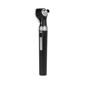
Otoscope
Otoscope Certified professional COMED
Otoscope Certified professional COMED
Otoscope Certified professional
Otoscope Certified professional
Otoscope Certified professional
Otoscope Certified professional
Otoscope Certified professional
Otoscope Certified professional
Otoscope Certified professional
In our "otoscope" booth, renowned vendors from around the world offer you a diverse range of high-quality otoscopes: from proven traditional models to cutting-edge technological innovations.
Designed for demanding professionals, the otoscopes showcased in our marketplace booth provide unparalleled diagnostic precision, ensuring that you can provide your patients with the highest level of care. Whether you need a robust otoscope for daily use in a busy clinic or a premium digital device with advanced imaging capabilities, you'll find it here.
Trust our certified partner merchants to provide you not only with the best quality but also impeccable customer service and fast, reliable delivery.
What is an otoscope?
An otoscope is an essential medical instrument used by healthcare professionals to examine the inner ear, tympanic membrane, and ear canal. It is a valuable tool for diagnosing conditions such as ear infections, earwax blockages, and other ear abnormalities.
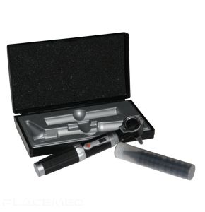
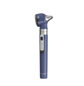
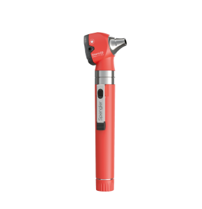
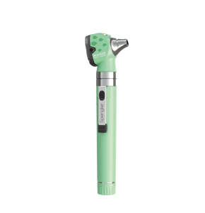
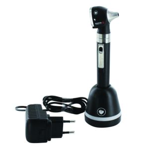
The otoscope consists of several key parts. First, there is the handle, which often also serves as the power source for the otoscope, typically in the form of batteries or an electrical connection. Next, there is the otoscope head, which contains a light source, often an LED or halogen bulb, that illuminates the ear for precise inspection. On this head, there is a magnifying lens, often rotatable, that helps enhance the visibility of ear structures.
Perhaps the most crucial component of the otoscope is the speculum, a cone-shaped piece that is inserted into the ear canal. Speculums are available in different sizes to fit various ages and sizes of ear canals.
The operating principle of the otoscope is quite simple. The light from the light source passes through the speculum and illuminates the ear canal and tympanic membrane, allowing the healthcare professional to clearly visualize these structures.
What is the history of the otoscope?
The otoscope has a rich and fascinating history!
Born in the first half of the 19th century, the otoscope was a response to the need for a tool that would allow physicians to examine the inside of the ear with unmatched precision. It started as a simple device, a tube with a light source, providing doctors with a coarse view of the inner ear.
Over time, the otoscope underwent a series of radical transformations. The advent of electricity in the 20th century brought a more reliable and brighter light source, thus improving the clarity of the view. Lens improvements allowed even more precise visualization of the inner ear. The design also modernized, with otoscopes becoming lighter, more compact, and more ergonomic, making them easier to use for healthcare professionals.
Today, the otoscope incorporates digital technology, offering high-resolution images that can be shared and examined in real time. From its initial design to its current form, the otoscope perfectly illustrates the constant innovation in the medical field.
What are the different types of otoscopes?
In the medical device market, there are different types of otoscopes that vary in their design and functionalities:
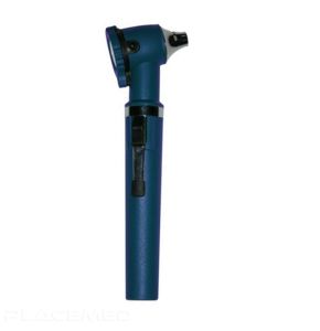
Pocket Otoscope
This type of otoscope is compact and portable, making it ideal for on-the-go use. It is often used by general practitioners and nurses to perform basic ear examinations. Pocket otoscopes are lightweight, easy to use, and equipped with a built-in light source to illuminate the ear.
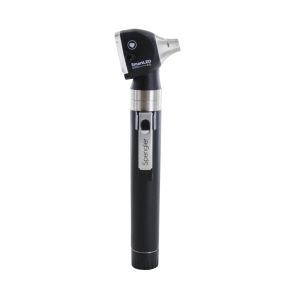
Fiber Optic Otoscope
These otoscopes use optical fibers to transmit light through the ear canal. They provide brighter illumination and better visibility compared to traditional otoscopes. Fiber optic otoscopes are commonly used by ear, nose, and throat specialists to perform more in-depth examinations and diagnose ear conditions.
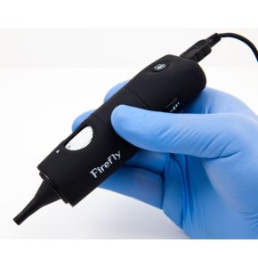
Video Otoscope
Video otoscopes are equipped with a built-in camera that allows visualization and capturing of images or videos of the ear. They provide enhanced visual documentation and can be useful for medical training and patient monitoring. Video otoscopes are often used in specialized clinics and healthcare facilities.
Pneumatic Otoscope
Also known as a pressure otoscope, this instrument is used to assess the mobility of the eardrum. It is equipped with a blowing device that applies gentle pressure to the eardrum to detect any abnormalities or obstructions. Pneumatic otoscopes are frequently used by ear specialists to evaluate hearing disorders and middle ear infections.
Specialized Otoscope
There are also specialized otoscopes designed for specific applications. For example, some otoscopes are equipped with accessories to perform audiometric tests or minor ear surgical procedures. These otoscopes are typically used by specialized healthcare professionals such as audiologists or otolaryngologists.
How to choose the right otoscope?
Choosing the right otoscope is crucial for healthcare professionals and even for home use. Whether you are a physician, medical student, or a concerned parent, the following points will help you understand the key aspects to consider when purchasing an otoscope.
- Image clarity: The otoscope should provide a clear and sharp image of the inner ear. Ensure to choose an otoscope with a good focusing lens and a powerful light source for better visibility.
- Otoscope size: The size and weight of the otoscope can impact your comfort and ease of use. Make sure to choose an ergonomic model that fits well in your hand.
- Light source: Quality lighting is essential for properly examining the ear canal. Fiber optic otoscopes are generally preferred as they provide shadow-free illumination of the examination area.
- Quality and durability: A high-quality otoscope will be made with durable materials and withstand the test of time. It may be slightly more expensive, but it is usually more cost-effective in the long run.
- Specula options: Specula are the tips that are inserted into the ear. They are available in different sizes and should be easy to change and clean.
 Francais

 Quote
Quote  Cart
Cart 