Ultrasound machines
18/11/2024 309
18/11/2024 283
18/11/2024 255
18/11/2024 282
18/11/2024 249
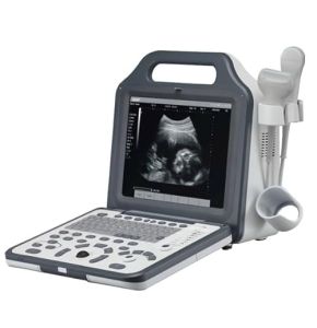
E-CUBE 15 Platinum Ultrasound System
Price on request18/11/2024 305
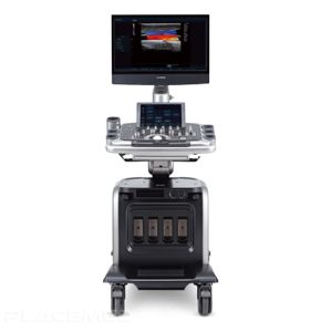
SONIMAGE MX1 Portable Ultrasound
Price on request18/11/2024 262
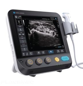
Ultrasound Machine on MyLab™X9 Platform
Price on request18/11/2024 260
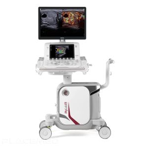
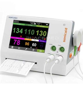
Sonicaid Team 3 I Fetal Monitor with Printer
Price on request18/11/2024 315
Ultrasound: An Indispensable Tool for Precise Diagnosis
In our "ULTRASOUND" booth, we offer a wide selection of high-quality devices, both portable and stationary, rigorously chosen for their exceptional performance quality. Ultrasounds are essential tools in modern medicine. They allow doctors to visualize the inside of the human body without surgical intervention. With the rise of portable ultrasounds, these devices have become even more accessible, offering increased flexibility and efficiency in many clinical situations.
Ultrasound is used in many medical specialties, from cardiology to obstetrics, and emergency medicine. It plays an important role in diagnosing, monitoring, and treating numerous medical conditions. Thanks to technological advancements, ultrasounds are now more efficient, more compact, and easier to use. This allows healthcare professionals to provide higher quality care to their patients.
What is Ultrasound and Why is it Important?
Ultrasound is a medical imaging technique that uses sound waves to create images of the inside of the body. Ultrasounds are high-frequency sound waves, inaudible to the human ear. By sending these waves into the body, they bounce off organs and tissues, creating echoes. The ultrasound machine captures these echoes and converts them into visible images on a screen.
This method is non-invasive, meaning it does not require incisions or the insertion of instruments into the body. Additionally, it does not use ionizing radiation, unlike X-rays or CT scans. This makes ultrasound particularly safe for patients, including pregnant women and children.
Ultrasound is important because it allows doctors to see in real-time what is happening inside the body. This helps to quickly diagnose many conditions, guide medical procedures, and monitor the progression of diseases. It is a valuable tool for providing precise and effective care.
How Do Ultrasounds Work?
Ultrasounds use a probe called a transducer. This probe emits ultrasounds when placed on the patient's skin. The ultrasounds penetrate the body and are reflected by different tissues and organs. The returning echoes are captured by the probe, which sends them to the ultrasound computer. The computer processes these signals and converts them into black and white or color images.
The obtained images can show the structure of organs, muscle movement, blood flow in vessels, and much more. Modern ultrasounds offer exceptional image quality, allowing the observation of very fine details. Some devices even offer three-dimensional (3D) or four-dimensional (4D) images, adding the dimension of time to view movements in real-time.
What Are the Advantages of Ultrasound?
- Non-invasive: No need for incisions or introduction of tools into the body, which reduces the risk of infection and pain for the patient.
- Safety: No use of ionizing radiation, making ultrasound safe for all patients, including pregnant women and children.
- Fast and Convenient: Ultrasound examinations can be performed quickly, often in just a few minutes, and results are available immediately.
- Versatile: Can be used to examine many parts of the body, including the heart, abdominal organs, muscles, joints, and blood vessels.
- Cost-effective: Ultrasound is generally less expensive than other imaging methods like CT scans or MRI.
What Are the Clinical Applications of Ultrasound?
Emergency Medicine and Intensive Care
In emergency medicine, ultrasound is a valuable tool. It allows doctors to quickly diagnose potentially life-threatening conditions. For example, in the case of trauma, ultrasound can detect internal bleeding, pneumothorax, or cardiac tamponade. This enables rapid decision-making to save the patient's life.
In intensive care, ultrasound is used to monitor the condition of critical patients. It helps evaluate heart function, detect fluids in the lungs or abdomen, and guide procedures such as catheter placement.
Anesthesia
Anesthesiologists use ultrasound to perform regional anesthesia with greater precision. By visualizing nerves and blood vessels, they can inject anesthetics exactly in the right location. This improves the effectiveness of anesthesia and reduces the risk of complications.
Other Applications
Ultrasound is also used in many other specialties:
- Cardiology: Echocardiography allows for the evaluation of heart function, detection of heart valve anomalies, or heart wall abnormalities.
- Obstetrics and Gynecology: Monitoring fetal development during pregnancy, evaluation of the uterus and ovaries.
- Gastroenterology: Study of the liver, gallbladder, pancreas, kidneys, and other abdominal organs.
- Orthopedics: Evaluation of muscles, tendons, ligaments, and joints to detect injuries or inflammations.
What Are the Innovative Technologies in Ultrasound?
Ultra-portable Ultrasounds
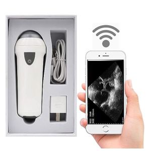
Technological advancements have led to the development of increasingly smaller and lighter ultrasounds. Some models fit in a pocket and can connect to a smartphone or tablet. This allows doctors to perform ultrasound examinations almost anywhere, whether in a clinic, at home, or even in the field.
These ultra-portable ultrasounds are particularly useful for care in rural or remote areas, where access to hospitals may be difficult. They also allow for rapid intervention in emergencies, thereby improving patient survival chances.
Artificial Intelligence
Artificial intelligence (AI) is increasingly integrated into modern ultrasounds. AI software can help interpret images, detect anomalies, and suggest possible diagnoses. This is particularly useful for less experienced doctors or when additional expertise is needed.
AI can also improve image quality by reducing noise and optimizing device settings. It can learn from accumulated data to provide increasingly accurate analyses over time.
Training in Virtual or Augmented Reality
Training doctors in the use of ultrasound is essential. Virtual reality (VR) and augmented reality (AR) technologies offer new possibilities for learning. With VR, doctors can train in a simulated environment, replicating real clinical situations without risk to patients.
AR can overlay useful information on the ultrasound image in real-time, such as annotations or positioning guides. This helps doctors interpret images more easily and improve their skills.
How to Train in Ultrasound?
Ultrasound is a powerful tool, but it requires adequate training to be used effectively. Many training programs are available for doctors and other healthcare professionals. These programs combine theoretical courses with supervised clinical practice.
Training can be tailored to the specific needs of each specialty. For example, a general practitioner can undergo basic training to perform simple examinations, while a cardiologist will require more advanced training in echocardiography.
Regular practice is essential to develop and maintain ultrasound skills. Workshops, seminars, and online courses can help professionals stay up to date with the latest techniques and technologies.
What Is the Impact of Ultrasound on Patient Care?
Ultrasound has a significant impact on the quality of care provided to patients. Here are some of the major advantages:
- Reduction of Diagnostic Time: By enabling rapid, on-site examinations, ultrasound accelerates the diagnostic process, which is crucial in emergency situations.
- Improvement of Care in Rural or Remote Areas: Portable ultrasounds bring advanced medical technology to areas where resources are limited.
- Reduction of Costs Related to Heavy Imaging Exams: Ultrasound is cheaper than CT scans or MRI, which helps reduce healthcare expenses.
- Guidance of Medical Procedures: Ultrasound helps doctors perform interventions with greater precision and safety.
- Improvement of Patient Satisfaction: Patients appreciate quick, non-invasive, and painless examinations.
In summary, ultrasound contributes to more effective, safer, and patient-centered medicine.
How to Maintain Your Ultrasound?
Regular maintenance of your ultrasound is essential to ensure its proper functioning: cleaning, updating, checking connections, etc.
- Cleaning After Each Use: Wipe the probe and device surfaces with appropriate products to prevent the buildup of dirt and bacteria.
- Checking Cables and Connections: Ensure that all cables are in good condition and properly connected to avoid malfunctions.
- Software Updates: Regularly install updates to benefit from the latest improvements and fixes.
- Proper Storage: Store the device in a dry and safe place when not in use.
- Preventive Maintenance: Schedule regular checks with a qualified technician to detect and resolve potential issues.
By following these tips, you ensure optimal performance and a long lifespan for your ultrasound.
Conclusion
Ultrasound is an essential technology in modern medicine. It provides healthcare professionals with a powerful tool to diagnose, treat, and monitor numerous medical conditions. With current innovations, ultrasounds are more accessible, more efficient, and easier to use than ever.
At Placemed, we are dedicated to providing healthcare professionals with the best equipment for their practice. Our selection of ultrasounds meets the varied needs of practitioners, whether for use in a clinic, hospital, or in the field.
Feel free to browse our online catalog and contact us with any questions. Together, let's improve the quality of care with cutting-edge technologies. Your success is our priority, and we are here to support you every step of the way.
 Francais